Homo sapiens: Eye, Ear and Mammalian Diversity
The Puzzle
Sense organs do not create perception alone.
They convert physical energy into patterned neural signals; the brain interprets those signals.
1. Systematic position
Chordata → Vertebrata → Mammalia → Primates → Hominidae → Homo sapiens.
2. Mammalian diagnostic characters
- hair;
- mammary glands;
- malleus, incus and stapes;
- dentary–squamosal jaw joint;
- muscular diaphragm;
- endothermy;
- four-chambered heart;
- heterodont dentition common;
- advanced parental care.
The most useful diagnostic combination is hair + mammary glands + three middle-ear ossicles.
Part I — Human Eye
3. Optical and neural design
The wall of the eye has:
- fibrous coat: sclera and cornea;
- vascular coat: choroid, ciliary body and iris;
- neural coat: retina.
Aqueous humor fills the anterior region; vitreous humor fills the posterior cavity.
4. Light pathway
According to the National Eye Institute:
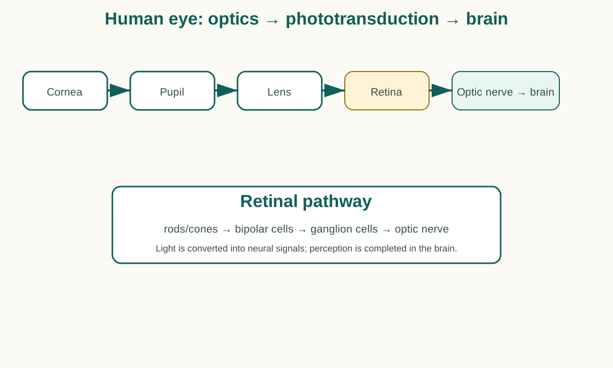
Light → cornea → pupil → lens → retina
→ electrical signals → optic nerve → brain
Cornea and lens focus light. The iris regulates pupil size.
5. Accommodation
For near objects, ciliary muscle contraction reduces zonular tension and the lens becomes more convex.
For distant objects, ciliary muscle relaxation increases zonular tension and flattens the lens.
6. Retina
The retina contains rods, cones, bipolar cells, ganglion cells and interneurons.
Simplified neural route:
rods/cones → bipolar cells → ganglion cells → optic nerve.
Rods
High sensitivity; dim-light vision.
Cones
Color and high-acuity vision; concentrated in the fovea.
The optic disc lacks photoreceptors and forms the physiological blind spot.
7. Vision mechanism
- cornea refracts light;
- pupil regulates entry;
- lens focuses;
- retina receives the optical pattern;
- photoreceptors transduce photons into electrical signals;
- retinal circuits process information;
- optic nerve carries ganglion-cell output;
- the brain creates visual perception.
The brain does not simply rotate an upside-down photograph; vision is active neural interpretation.
Part II — Human Ear
8. Outer, middle and inner ear
Outer ear
pinna + auditory canal.
Middle ear
tympanum + malleus + incus + stapes + Eustachian tube.
Inner ear
cochlea + vestibule + semicircular canals.
9. Hearing mechanism
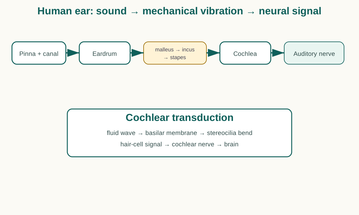
NIDCD describes the sequence:
Sound wave
→ ear canal
→ tympanic membrane
→ malleus → incus → stapes
→ oval window
→ cochlear fluid wave
→ basilar membrane
→ hair-cell stereocilia
→ electrical signal
→ auditory nerve
→ brain
The ossicles improve mechanical transfer from air to cochlear fluid.
10. Cochlea and pitch
The organ of Corti sits on the basilar membrane.
Hair-cell stereocilia bending produces receptor signals.
The basilar membrane is tonotopic:
- base → higher frequencies;
- apex → lower frequencies.
11. Balance
Semicircular canals detect angular acceleration. Utricle and saccule detect linear acceleration and head position.
12. Common sensory logic
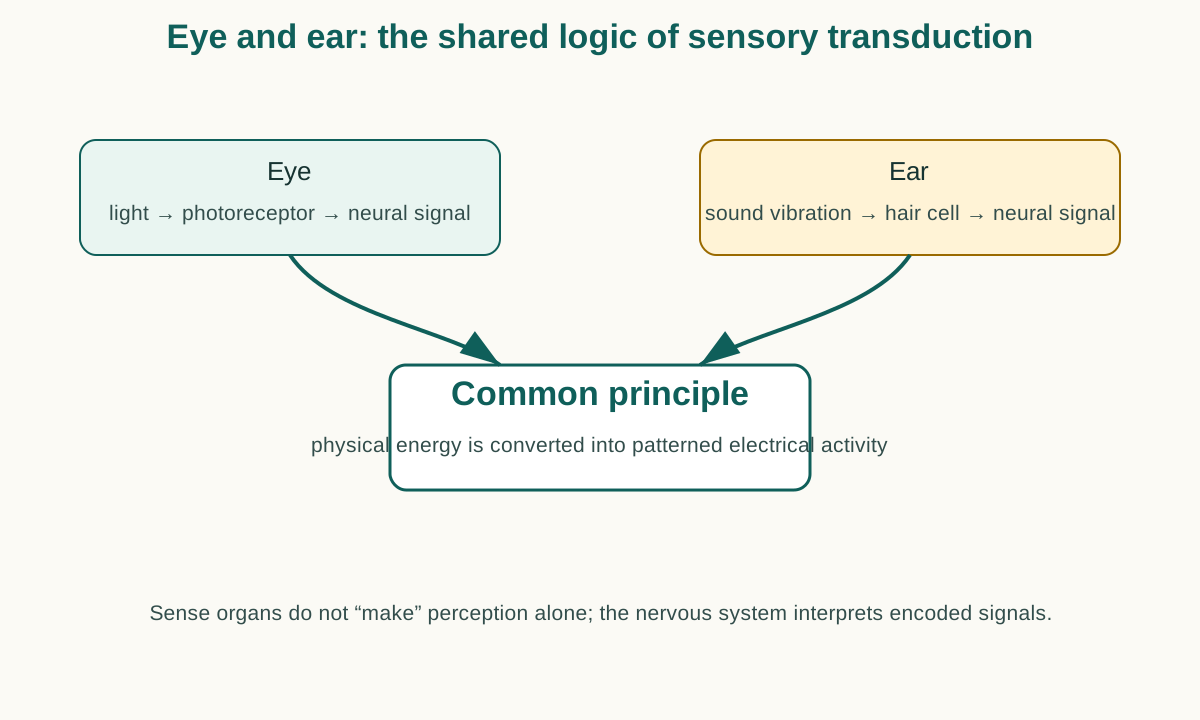
| Eye | Ear |
|---|---|
| light | sound/mechanical energy |
| rods/cones | hair cells |
| phototransduction | mechanotransduction |
| optic nerve | cochlear/vestibular nerve |
| visual perception | auditory/balance perception |
13. Mammalian diversity
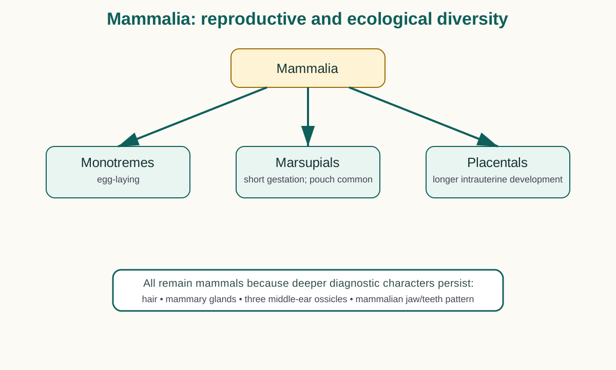
Monotremes
Egg-laying mammals such as platypus and echidnas.
Marsupials
Shorter gestation; extremely immature young; a pouch is common in many species.
Placentals
Prolonged intrauterine development supported by a complex placenta.
14. Aquatic mammals
Whales, dolphins, seals and manatees remain mammals because deeper diagnostic characters outweigh aquatic streamlining.
15. Apes and humans
Humans are apes in modern taxonomy. Hominoids lack an external tail and share mobile shoulders and multiple cranial/skeletal characters.
16. Bengal tiger
Panthera tigris tigris shows mammalian traits combined with carnivoran specializations such as large canines, carnassials, claws and predatory sensory systems.
17. Section A — one-mark capsule
Retina: neural light-sensitive tissue lining the back of the eye.
Optic nerve: ganglion-cell axons carrying visual signals to the brain.
Auditory ossicles: malleus, incus and stapes.
Organ of Corti: cochlear sensory epithelium on the basilar membrane.
Diastema: toothless gap between tooth groups.
18. Section B — vision
Cornea and lens focus light on the retina. Rods and cones convert light to electrical signals. Retinal circuits process the signals and the optic nerve carries output to the brain.
19. Section B — hearing
Sound vibrates the tympanum, ossicles transmit motion to the oval window, cochlear fluid moves the basilar membrane, hair cells transduce vibration and the cochlear nerve carries the signal to the brain.
20. Section C — eye
Describe the three coats, lens and humors, retina and optic nerve; then trace light focusing and phototransduction. Include a labelled cross-section.
21. Section C — ear
Describe outer, middle and inner ear, then trace the vibration-to-neural pathway. Include a labelled diagram.
22. Common mistakes
- calling the pupil a black tissue;
- making the lens the sensory receptor;
- treating the retina as a passive screen;
- attaching stapes to the round window;
- making semicircular canals the main hearing organ;
- assuming adult human hair cells regenerate freely;
- classifying whales as fishes.
23. Quick revision
| System | Sequence |
|---|---|
| vision | cornea → pupil → lens → retina → optic nerve → brain |
| hearing | canal → eardrum → ossicles → cochlea → nerve → brain |
| retina | rods/cones → bipolar → ganglion |
| ossicles | malleus → incus → stapes |
| balance | vestibule + semicircular canals |
| mammal hallmark | hair + mammary glands + 3 ossicles |
24. Research-backed update notes
National Eye Institute material supports the cornea/lens → retina → optic nerve sequence. NIDCD describes the tympanum → ossicles → cochlea → hair-cell → nerve pathway. Mammalian identity depends on diagnostic anatomy, not a single ecological or reproductive feature.
References for Course Authors / Verification
- National University Zoology syllabus, paper 223101.
- National Eye Institute, How the Eyes Work.
- NIDCD, How Do We Hear?
- Standard human anatomy and physiology texts.
- Modern mammalian taxonomy references.
Course synthesis
Chordate body plan
→ tunicate metamorphic specialization
→ cephalochordate retained body plan
→ living jawless vertebrates
→ cartilaginous jawed fishes
→ bony-fish gill efficiency
→ amphibian land–water compromise
→ reptilian amniotic independence
→ avian high-performance respiration
→ mammalian sensory specialization
This is a comparative scaffold, not a literal ladder of modern species.
End of Core Lecture Series
Next layer: comparative-system matrix, past questions and exam intelligence.